Sonography

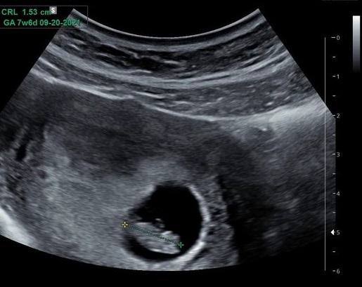
Obstetric ultrasound, also known as prenatal or pregnancy ultrasound, uses high-frequency sound waves to produce images of a developing embryo or foetus. The procedure also monitors the health of the mother’s uterus, ovaries, and the blood flow through the umbilical cord to the placenta

Reasons for an Obstetric Ultrasound

Your doctor may refer you for an obstetric ultrasound to:

  • Confirm fertilization, implantation, and the presence of an embryo
  • Identify whether it is a single or multiple pregnancy
  • Estimate the fetus’s age and the due date
  • Evaluate the position of the fetus and placenta in relation to the birth canal
  • Check the amount of amniotic fluid around the fetus
  • Monitor fetal development
  • Check for congenital disabilities, hereditary conditions, or abnormalities

First-Trimester Combined Screening (FTS)

FTS is a test for chromosome conditions and congenital disabilities. It is available to all women between 11 and 13 weeks plus six days of pregnancy. With FTS, two measurements are used, together with your age, to estimate your chance of having a baby with Down syndrome, trisomy 18, or trisomy 13. The first measurement is the nuchal translucency (NT) measurement, taken by ultrasound, and the second is a blood test.

Nuchal Translucency (NT) Measurement

NT ultrasound exams are performed between 11 weeks and 13 weeks plus six days of pregnancy. During an NT exam, your sonographer will measure the fluid at the back of the Fetuses neck (called nuchal translucency). All Fetuses have some fluid here. Fetuses with Down syndrome are more likely to have an increased amount of fluid.

The NT ultrasound also:

  • Confirms that the baby has a consistent heartbeat
  • Confirms your dates
  • Diagnoses a multiple pregnancy
  • Checks for other congenital disabilities

The Maternal Blood Test

The maternal blood test measures two substances, PAPP-A and free beta-hCG, that are usually found in the blood of all pregnant women. When Down syndrome is present, the levels of free beta-hCG and PAPP-A tend to be out of the expected normal range.

What is Anomaly Scan?

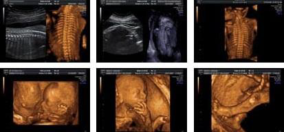
Anomaly Scan or mid-pregnancy scan is an ultrasound scan done between the 18th and 21st week of pregnancy to take a closer look at the baby and the womb (uterus) and to have an idea where the placenta is lying.

The scan aims to look for any major physical abnormalities in the growing baby. The scan can be as a dating scan where a black and white 2-dimensional (2-D) image is produced which gives the side-view of the baby in the womb. This image shows the baby’s face and hands at 20 weeks and gives the healthcare specialist (a sonographer) an idea of what is going inside. This can be undoubtedly exciting!

Why is Anomaly Scan done?

The mid-pregnancy anomaly scan is done for checking any physical abnormalities in the growing baby. Although it can’t pick up every problem, it gives the healthcare specialist (a sonographer) an idea about the baby’s bones, heart, brain, spinal cord, face, kidneys and abdomen and allows the healthcare specialist identify the following conditions (some of which are very rare):

  • Anencephaly
  • Diaphragmatic hernia
  • Gastroschisis
  • Exomphalos
  • Open spina bifida
  • Bilateral renal agenesis
  • Lethal skeletal dysplasia
  • Edwards’ syndrome or T18
  • Patau’s syndrome or T13
  • Cleft lip
  • Serious cardiac abnormalities

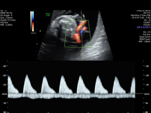
Doppler ultrasound uses sound waves to detect the movement of blood in vessels

Indications

Umbilical Doppler assessment is indicated in scenarios where there is a risk of foetal growth restriction or poor perinatal outcome. It is also used to stage twin-twin transfusion

Doppler ultrasound evaluation of the fetoplacental circulation is not indicated in low-risk pregnancies .

Maternal conditions

  • diabetes mellitus
  • chronic kidney disease
  • hypertension
  • prothrombotic states

Pregnancy-related conditions

  • suspected IUGR
  • previous pregnancy with IUGR or foetal death in utero
  • decreased fetal movement
  • oligohydramnios
  • polyhydramnios
  • multifetal pregnancy

What is fetal echocardiography?

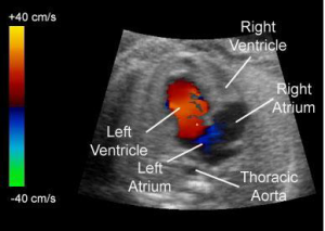
Fetal echocardiography is a test similar to an ultrasound. This exam allows your doctor to better see the structure and function of your unborn child’s heart. It’s typically done in the second trimester, between weeks 18 to 24.

The exam uses sound waves that “echo” off the structures of the fetus’s heart. A machine analyzes these sound waves and creates a picture, or echocardiogram, of their heart’s interior. This image provides information on how your baby’s heart formed and whether it’s working properly.

It also enables your doctor to see the blood flow through the fetus’s heart. This in-depth look allows your doctor to find any abnormalities in the baby’s blood flow or heartbeat.

When is fetal echocardiography used?

Not all pregnant women need a fetal echocardiogram. For most women, a basic ultrasound will show the development of all four chambers of their baby’s heart.

Your OB-GYN may recommend that you have this procedure done if previous tests weren’t conclusive or if they detected an abnormal heartbeat in the fetus.

You may also need this test if:

  • your unborn child is at risk for a heart abnormality or other disorder
  • you have a family history of heart disease
  • you’ve already given birth to a child with a heart condition
  • you’ve used drugs or alcohol during your pregnancy
  • you’ve taken certain medications or been exposed to medications that can cause heart defects, such as epilepsy drugs or prescription acne drugs
  • you have other medical conditions, like rubella, type 1 diabetes, lupus, or phenylketonuria
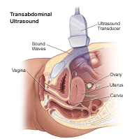
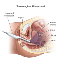
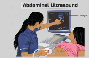
The examination can be performed by transabdominal ultrasonography, generally with a full bladder which acts as an acoustic window to achieve better visualisation of pelvis organs, or by transvaginal ultrasonography with a specifically designed vaginal transducer. Transvaginal imaging utilises a higher frequency imaging, which gives better resolution of the ovaries, uterus and endometrium

Indications:

  • to assess pelvic organs,
  • to diagnose acute appendicitis
  • to diagnose and manage gynecologic problems including endometriosis, leiomyoma, adenomyosis, ovarian cysts and lesions,
  • to identify adnexal masses, including ectopic pregnancy,
  • to diagnose gynecologic cancer
  • in infertility treatments to track the response of ovarian follicles to fertility medication (i.e. Pergonal). However, it often underestimates the true ovarian volume.

Abdominal and pelvic ultrasound is a type of imaging test.

What is the difference between an abdominal and pelvic ultrasound?

Doctor considers abdominal ultrasound a type of pelvic ultrasound because it evaluates tissues inside the pelvis (hip bones). Other types of pelvic ultrasound include transvaginal ultrasound and rectal ultrasound.

What body parts does an abdominal ultrasound evaluate?

Your provider orders ultrasound evaluation of specific areas of your abdomen. A right upper quadrant ultrasound examines three organs of the digestive system:

  • Liver.
  • Pancreas.
  • Gallbladder.

A complete abdominal ultrasound examines those three organs and adds the:

  • Kidneys.
  • Spleen.
  • Bladder.
  • Abdominal blood vessels (such as the inferior vena cava and the aorta).